 Reproduced, with permission, from:
Longstreth, J. D., ed. 1987. Ultraviolet radiation and melanoma-with a special focus on assessing the risks of stratospheric ozone depletion. Vol. 4, Appendix A of Assessing the risk of trace gases that can modify the stratosphere. Washington, D.C.: U.S. Environmental Protection Agency.
Reproduced, with permission, from:
Longstreth, J. D., ed. 1987. Ultraviolet radiation and melanoma-with a special focus on assessing the risks of stratospheric ozone depletion. Vol. 4, Appendix A of Assessing the risk of trace gases that can modify the stratosphere. Washington, D.C.: U.S. Environmental Protection Agency.
STEROID HORMONES AND MALIGNANT MELANOMA
Evidence for a relationship between the biological behavior of melanoma and steroid hormone action has been observed in several areas of research. These observations include differing survival prognoses favoring females over males, the rarity of the tumor in prepubescent children, the improved survival for postmenopausal and multiparous women, and the increased melanoma incidence among 30- to 50-year-old women. Effects of pregnancy and exogenous hormones on growth and development of melanomas have also been shown. Estrogen receptors have also been observed in human melanomas (McCarty et al. 1980).
Effects of Endogenous Hormones
Several studies indicate that sex hormones may influence melanocyte activity and the natural history of malignant melanoma. Based on the observed rapid increase in mole counts in both sexes during puberty, MacKie et al. (1985) suggested the presence of a hormonal influence on the pigment-producing activity of nevi. The authors suggested that this could result from either a new appearance and proliferation of pigment-producing nevi or the activation of the melanin-producing enzyme pathway of pre-existing, inactive, non-pigment-producing nevi.
Hodgins (1983) noted that the pigmentation changes of genital and areolar skin at puberty and in pregnancy suggest that gonadal hormones influence at least some populations of melanocytes. Greene et al. (1985) stated that they counsel high-risk family members (those with dysplastic nevi) to pay particular attention to nevi during periods of hormonal flux (i.e., puberty and pregnancy).
Several epidemiologic studies have indicated that the observed sex differences in melanoma incidence and mortality could be related to hormonal differences. Hodgins (1983) concluded, however, that there is no clear evidence linking these differences to levels of steroid hormones. For example, the prognosis for men with malignant melanoma is worse than for women. Hodgins cited Shaw et al. (1978), who concluded that better survival resulted at least in part from the earlier stage at presentation and prognostically more favorable sites among women. However, when male patients were matched to female patients by age, and size and thickness of lesion, the female survival advantage among premenopausal stage I patients compared to matched male patients persisted. The survival advantage was much less for postmenopausal women compared to matched male patients. The results, Hodgins noted, supported the concept of a barrier to tumor metastasis in premenopausal women.
The observation of higher melanoma incidence and mortality rates among reproductive and menopausal-aged women than among men of the same age in the British Isles led Lee and Storer (1980) to suggest that a hormone-dependent variant of melanoma may account for the difference. The authors conducted a descriptive comparison of WHO mortality data from eight European countries and Office of Population Censuses and Surveys incidence data from England and Wales. Age-specific female-to-male mortality ratios indicated higher female mortality rates relative to male rates in the British Isles from 1955 to 1974, whereas the reverse was observed for Australia, North America (the U.S. and Canada), Japan, and Scandinavia (Denmark, Norway, Sweden, and Finland). Lee and Storer (1980) did not offer an explanation for the higher British Isles mortality ratios. They noted, however, that a similar pattern of age-specific sex ratios (i.e., risks for females compared with males peaked in the latter half of reproductive life, and decreased or leveled off in middle age) was observed in these different populations.
Excess female mortality and incidence rates relative to those in males in the British Isles was greatest from ages 30-44. The elevated sex ratio from ages 30-49 did not change from 1950 to 1974. Lee and Storer (1980) suggested that the low rates for malignant melanoma in the British Isles, compared to those for Australia and New Zealand, permitted the observation of a hormone-dependent variant of melanoma in the British Isles.
Lee and Storer (1982) analyzed age-specific changes in the sex ratio of malignant melanoma for several countries of Europe, North America, Australia, and Japan using WHO mortality data. The female/male mortality ratio was less than 1.0 for all countries examined, in contrast to the female excess in the British Isles (Lee and Storer 1980). In each of these populations, however, female/male sex ratios peaked during the reproductive years and declined in middle age. The authors tested whether an interaction of sex and age on CMM mortality rates could have been produced by birth cohort effects and sex differences in incidence and mortality. An examination of the data by 5-year birth cohort intervals indicated a persistent increased female risk in the reproductive years. Using a mathematical model to separate age and cohort effects, Lee and Storer observed that interaction terms for age and sex and year of birth and sex were both significant. They concluded that the specific variation in female-to-male ratios may reflect the same biologic progress that underlies changes in melanoma survival in relationship to childbearing (e.g., decreased survival among pregnant women).
Holman et al. (1984) conducted a case-control study on 276 female melanoma patients identified in the West Australia Lions Melanoma Research Project from 1980 to 1981. Two hundred and seventy-six age- and electoral-subdivision-matched controls were selected from the Australian Commonwealth Electoral Roll, and a few from public school student rolls. The authors observed no consistent evidence of a relationship of incidence rates of different histogenic types with age at menarche, duration of menstrual life, or number of pregnancies of over 20 weeks' duration.
Effects of Pregnancy
In a brief communication in Lancet, an anonymous writer (Anon 1971) stated that while the relationship of pregnancy and melanoma used to be a matter of debate, pregnant women run the same risk of developing melanoma as non-pregnant women, and tumor behavior is similar in the two groups. Scattered evidence has indicated, however, that pregnancy can activate metastatic disease in a previously treated melanoma, or increase its growth rate (Lee and Storer 1982; McCarty et al. 1980).
In a study by Foucar et al. (1985), 86 pregnant white patients visiting the obstetrics clinic of the University of Iowa Hospitals and Clinics between June and August 1982 permitted the removal of 128 nevi for study. Fifty-one non-pregnant female controls and 50 male controls were obtained from the Department of Pathology files of the University of Iowa Dermatology Clinic. Controls were excluded if they were not between the ages of 16-39. In addition, controls with evidence of atypical nevi were not included. Case and control nevi were compared using a graduated scoring system for atypia ranging from 1 to 16. The 128 nevi from pregnant patients did not include any lesions with sufficient atypia to suggest malignancy. The histopathologic features among cases' nevi were identical to those seen in the male controls' nevi. However, based on small but noticeable differences among histopathologic "activation" measures between cases' nevi and nevi from female controls, Foucar et al. (1985) suggested that mild changes in some nevocellular nevi may occur during pregnancy. The authors noted, however, that a potential bias in selecting controls may have resulted from elimination of controls with clinical features potentially associated with histopathologic atypia.
Most epidemiologic investigations on pregnancy and melanoma have focused on differences in survival of melanoma patients by pregnancy status at or near the time of diagnosis. In an analysis of survival data for female melanoma patients of childbearing age, White et al. (1961) observed that pregnancy did not have an adverse effect on survival even after stratifying by age and stage of disease. The study population consisted of 18 women seen at Stanford Hospital and 53 women from the California Tumor Registry aged 15 to 39 years at CMM diagnosis. The patients were divided into a pregnant group (N=30) (those pregnant within 1 year before and 5 years after diagnosis), a non-pregnant group (N=31), and a pregnancy-undetermined group (N=10). Five-year survival rates were examined for the three groups and indicated a higher survival rate for pregnant (73 percent) than non-pregnant patients (55 percent), although the difference was not significant due to the small sample size. To test the robustness of the observed differences between the pregnant and non-pregnant groups, a range of extreme assumptions concerning the makeup of the pregnancy-undetermined group was applied. The results indicated that within the limits of the data, 5-year survival rates among pregnant women were similar to, or greater than, rates among non-pregnant women. Within three age groups (under 20, 20-29, 30-39), survival rates were still equal to or more favorable than rates for non-pregnant women. When analyzed by stage of disease, White et al. (1961) observed that among those with localized disease (25 cases pregnant, 21 non-pregnant), 5-year survival rates were slightly higher among pregnant patients (88 percent) than non-pregnant patients (81 percent). It should be emphasized that the number of women in this study was small and may not have been adequate for the detection of differences by pregnancy status. The authors concluded that, based on their data, pregnancy did not appear to have a deleterious effect on survival.
Shiu et al. (1976) found lower 5-year survival rates in pregnant women who had experienced activation of a lesion during a previous pregnancy, based on a survival analysis of 251 female cases ages 15-45 who received treatment at the Memorial Sloan-Kettering Cancer Center from 1950 to 1969. Cases were selected if accurate recorded data on pregnancy at the time of admission were available. The authors observed no significant difference in 5-year survival rates for Stage I patients (N=165) between nulliparous, parous non-pregnant, and pregnant women. Among 86 Stage II patients, however, significantly lower survival rates (p<=0.05) were observed for pregnant patients (29 percent) and parous patients who had lesion activation in a previous pregnancy (22 percent) as compared with nulliparous patients (55 percent) and other parous patients (51 percent) combined. Age differences did not account for the differences in 5-year survival rates. Shiu et al. (1976) concluded that the differences in survival rates and frequency of symptoms in Stage II patients (e.g., bleeding, ulceration, and elevation of lesion) "strongly suggest an adverse influence of pregnancy on women with stage II melanoma."
Hodgins (1983) has noted, however, that suggestions of adverse effects of pregnancy upon malignant melanoma have not been supported by more recent epidemiologic studies. Elwood and Coldman (1978) observed that 5-year survival rates did not differ among 254 ever-pregnant and 51 never-pregnant melanoma patients. The study population was comprised of 305 consecutive melanoma patients seen in Vancouver and diagnosed between 1960 and 1976. The authors noted that their results differed from those of Hersey et al. (1977) who studied 443 consecutive female patients seen in Sydney from 1961 to 1971. Substantially better survival rates for ever-pregnant women were reported; the largest difference in survival was for women over 50 with survival rates of 73 percent (ever-pregnant) and 53 percent (never-pregnant). After restricting their series to cutaneous lesions (89 percent of the total) and adjusting for stage at diagnosis, Elwood and Coldman (1978) still did not observe any association between survival and pregnancy history. Elwood and Coldman concluded that the inconsistency between their results and those of Hersey et al. argued against the hypothesis of improved survival among ever-pregnant melanoma patients.
Houghton et al. (1981) compared 3- and 5-year survival rates among female melanoma patients aged 15 to 40 years of age using data obtained from the Connecticut Tumor Registry from 1950-1954, 1960-1964, and 1970-1974. The study included 12 patients diagnosed during pregnancy (cases) and 175 patients not pregnant at the time of diagnosis. Each case was matched with two non-pregnant patients according to age, anatomic site, and stage of disease at diagnosis. No differences in survival between the two case groups were noted. The 3-year survival rate was 65 percent among pregnant patients versus 67 percent among matched non-pregnant patients; the 5-year survival rate was 55 percent among pregnant versus 58 percent among matched non-pregnant cases. The expected number of pregnant women among the 187 patients reviewed, estimated from Connecticut livebirth rates, was 13.3, compared to the observed 12 pregnancies, suggesting that melanoma incidence did not substantially increase during pregnancy. Houghton et al. (1981) noted that survival rates were significantly lower among pregnant cases when age, anatomic site of primary lesion, and stage at diagnosis were not considered.
Effects of Exogenous Hormones
Several studies have examined the possible relationship between oral contraceptive (OC) and other hormone use and melanoma. Beral et al. (1984) concluded, based on available epidemiologic evidence, that "while oral contraceptives and other exogenous sex hormones are clearly not major determinants of melanoma, the accumulating evidence suggests that they may increase the risk of disease."
Jelinek (1970) observed that pigmentary changes (melasma) caused by oral contraceptives suggest that estrogen or progesterone could control skin melanin. According to Jelinek, estrogens stimulate melanocytes and progesterone causes the pigmentation to spread, indicating that the total amount of both hormones could increase melasma incidence. Jelinek (1970) stated, however, that the putative relationship between malignant melanoma and OC use could be adequately explained by chance. MacKie et al. (1985) observed that neither pregnancy nor OC use stimulate development of new moles, although some pre-existing moles have been found to temporarily darken during pregnancy. Hodgins (1983) noted that in spite of the observed pigmentary changes associated with OC use (attributable mainly to estrogens), there is little evidence to suggest any link between CMM and the contraceptive pill.
Lederman et al. (1985), in a prospective study of 289 Caucasian female Stage I melanoma patients, conducted a multivariate analysis to examine the effects of prior estrogen and progesterone use on tumor characteristics and survival. The cases were consecutively evaluated and prospectively entered into a natural history study at the Massachusetts General Hospital and New York University. Hormone users presented with thinner tumors than nonusers; 76 percent of OC users and 64 percent of menopausal estrogen (MPE) users had primary tumors less than 1.69 mm, as compared with 58 percent of nonusers. Users of OCs in the year prior to CMM diagnosis had significantly thinner tumors than nonusers (p<=0.01) in the year before diagnosis. Univariate analysis showed that exogenous hormone use was not associated with increased risk of death from CMM. Life table analysis revealed slightly greater 5- and 9-year survival rates in hormone users. Nine-year survival rates were 90 percent for OC users, 87 percent for MPE users, and 81 percent for non-users. The finding of thinner tumors in hormone users may have explained their apparently more favorable survival. The fact that estrogen users had thinner tumors may have been due to a direct effect of estrogens, the tendency of hormone users to seek medical attention sooner than non-users, or that hormone users tend to be under closer medical surveillance than non-users.
Lee and Storer (1982) noted that in the British Isles population, the elevated female-to-male melanoma incidence ratio did not change over successive 5-year periods during a 25-year span from 1951 to 1975. These data suggested that the large-scale introduction of OCs did not produce the elevated ratio.
Using the same methodological approach as Lee and Storer (1982), Stevens et al. (1980) examined incidence data from Connecticut (1935-1974), Denmark (1943-1972), and Finland (1953-1974), and mortality data from the United States (1951-1975), Canada (1951-1975), and England and Wales (1951-1975). The authors observed that there was no perturbation in female melanoma incidence rates relative to male incidence rates at or after the introduction of oral contraceptives. On the basis of these descriptive data, Stevens et al. (1980) argued that there is no association between OC use and malignant melanoma since, if there were an association, male incidence and mortality rates would not be a constant multiple of female rates over an extended period of study.
Beral et al. (1984) conducted a case-control analysis of the effect of OC and hormone use, and pregnancy history on risk of melanoma. The cases consisted of 287 white women aged 15-84 years who were seen at the Sydney Hospital. General population controls were matched by age and area of residence to cases diagnosed between 1974 and 1978 ("old" cases). Controls selected from hospital inpatients were matched to "new" cases (those interviewed between 1978 and 1980) by age only. Case-control comparisons indicated that women with melanoma were more likely to have taken oral contraceptives for long periods of time. A consistently increased risk of melanoma was only observed in those who had begun OC use at least 10 years before diagnosis and whose use continued for 5 or more years relative to non-users (relative risk [RR]=1.5; 95% CI 1.03-2.14). This elevated risk persisted after controlling for reported hair and skin color, frequency of moles on body, place of birth, and measures of sunlight and fluorescent light exposure. Socioeconomic status, which has been associated with CMM (see Chapter 11), was not controlled for in this study. If there were an association between socioeconomic status and OC use, the failure to control for socioeconomic status may confound the observed results. Beral et al. (1984) also observed that cases were more likely than controls (but not significantly) to have used hormones to regulate periods (RR=1.9), to have received hormonal replacement therapy (RR=1.4), and to have been given hormone injections to suppress lactation (RR=1.4).
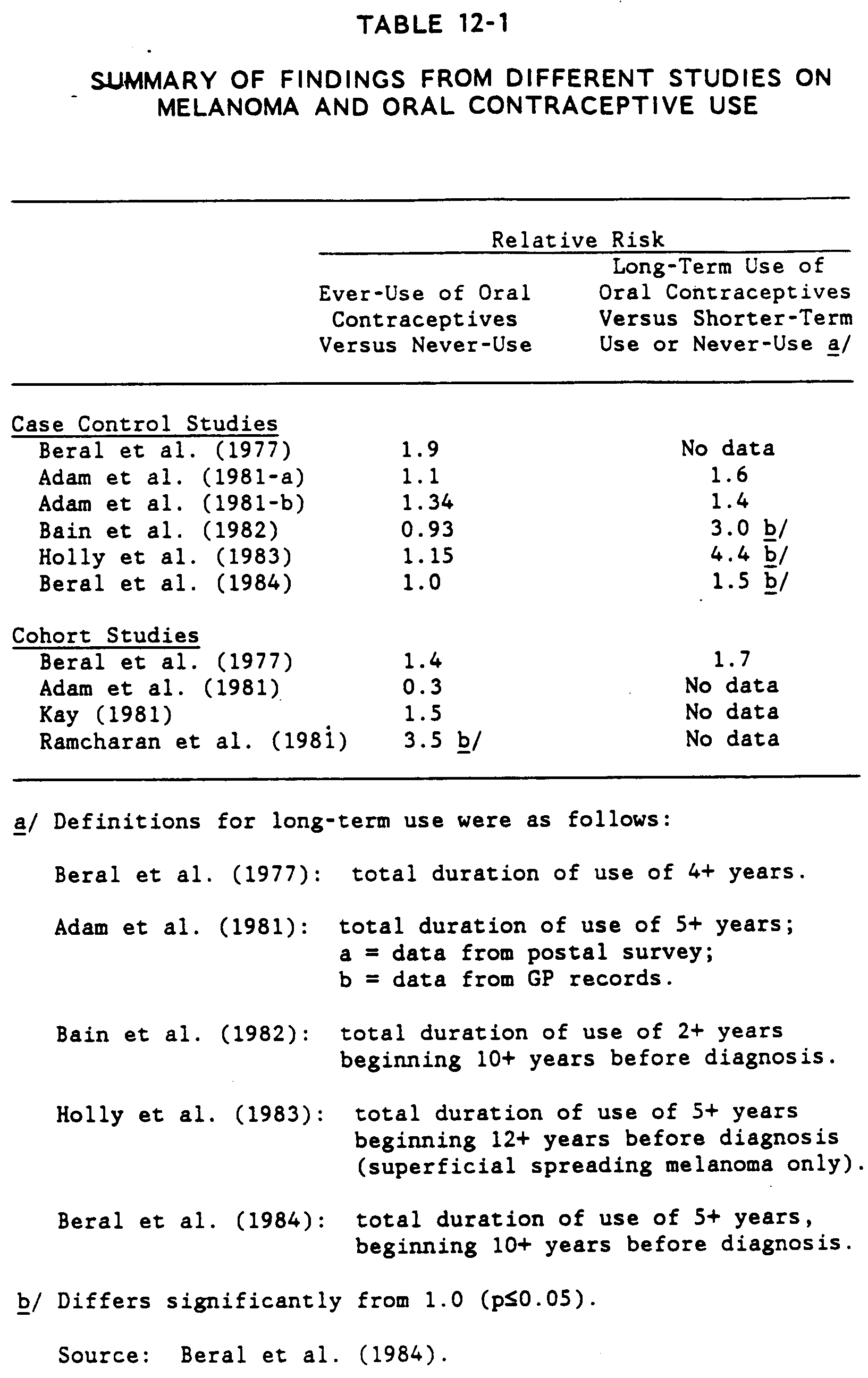
Based on conclusions from several studies, Beral et al. (1984) stated that while most studies reported weak or no associations of CMM to ever-use of OCs, the five studies examining data on prolonged OC use found increased risks (not always statistically significant) associated with long-term pill use. As shown in Table 12-1, relative risk estimates for long duration of OC use are in the range of 1.4 to 4.4. These relative risks are of the same order of magnitude as those for recognized pigmentary risk factors such as red or blonde hair and fair skin. It should be kept in mind, however, that some of these results may be confounded by socioeconomic status.
Endocrine Therapy
In breast cancer, the presence of specific estrogen receptors in a tumor has provided an indication of responsiveness to endocrine therapy. Several studies have searched for hormone receptors in malignant melanomas in the hope of identifying patients who might respond to hormone treatment. Studies have shown that steroid receptors are a necessary, if not sufficient, requirement for steroid-hormone responsiveness in target tissues (Fisher et al. 1976).
Occurrence of Hormone Receptors. Based on a review of 14 published studies, Hodgins (1983) concluded that melanomas generally contain low estrogen receptor concentrations, below those considered significant for predicting hormone responsiveness for breast cancer. Ellis et al. (1985) noted, however, that several studies describing hormonal receptors in human malignant melanoma have found that 0-78 percent of melanomas have detectable levels of estrogen receptors, and from 0-100 percent of melanomas have detectable levels of progesterone receptors.
McCarty et al. (1980) examined tumors from 20 patients aged 23 to 80 years hospitalized at the Duke Comprehensive Cancer Center. No restriction was made concerning age, sex, race, or menstrual status of the patients included in the study. Seven of the 20 tumors showed high affinity estrogen binding of more than 3 fmol/mg cytosol tissue protein by dextran-coated charcoal analysis (DCCA). No relationship was observed between estrogen binding and age, sex, menstrual status, or parity. No evidence of high affinity progesterone binding was observed in any of the 20 tumors. The authors cautioned, however, that their results also supported the hypothesis that the enzyme tyrosinase may mimic estrogen receptor binding. The possibility that steroids may interact with nonreceptor proteins such as tyrosinase gives less credence to the specificity of estrogen binding to receptors in melanoma skin tumors.
Rumke et al. (1980) conducted hormone-receptor assays on 21 metastatic tumors from 17 male patients, and 22 metastatic tumors from 17 female melanoma patients. Estrogen and androgen receptors were detected in 7 out of 31 cutaneous metastases. No relationship was observed between estrogen receptor and sex, age, androgen receptor, or prognosis. The study showed that estrogen and androgen receptors can be present in some melanoma metastases but at levels generally too low to be considered of relevance to endocrine treatment.
Creagan et al. (1980) assayed 38 tumor specimens from 34 melanoma patients for cytoplasmic estrogen receptors (ER) by the dextran-coated charcoal method. Only 4 of the 34 patients (12 percent) had detectable ERs, leading the authors to conclude that the chemical usefulness of the ER assay in melanoma is probably limited.
Fisher et al. (1976) analyzed biopsies from 35 malignant melanoma patients and found 16 (46 percent) with cytoplasmic receptors for estrogen. Equal percentages were observed for males and females. Using the Scatchard technique, the authors observed a straight line plot suggesting that estradiol was binding to a single class of high affinity, limited capacity receptor sites.
Ellis et al. (1985) investigated a related topic: whether hormone-receptor binding in melanocytic lesions could be indicative of a potential for malignancy. Estrogen and progesterone binding was examined in 22 melanocytic lesions from 14 patients with the dysplastic nevus syndrome and in 21 patients with acquired intradermal nevi using a fluorescent hormone binding technique. Large amounts of both estrogen and progesterone binding were seen in nevi from patients with dysplastic nevus syndrome, while most of the acquired intradermal control nevi were negative for binding. The authors concluded that positive estrogen and progesterone binding in melanocytes from patients with dysplastic nevi may correlate with clinical lesion changes during times of normal hormonal change (e.g., puberty, pregnancy, and OC use). The authors noted that the predictive value of estrogen and progesterone receptors in melanocytic lesions for malignancy remains unproven.
Effects of Endocrine Treatment. In a 1983 review article, Hodgins (1983) concluded that estrogens and anti-estrogens appeared to be useful in treatment of some melanomas, but more work was needed to identify patients likely to respond. There was little evidence concerning the effects of castration, androgen, or antiandrogens in the treatment of melanoma. Hodgins cautioned that glucocorticoids and retinoids, while able to inhibit the growth of melanoma cells, have widespread negative health effects. Finally, Hodgins stated that assays of steroid sex hormone receptors in melanomas appeared to offer little help in selecting patients for endocrine treatment. While premalignant melanocytic lesions and early-stage tumors might be estrogen-dependent, Hodgins suggested that as tumors spread, hormone-resistant variants become established. Tumors could then contain a mixed population of hormone-responsive (staining) and hormone-insensitive (nonstaining) cells.
Most hormonal treatments for CMM involve administration of the anti-estrogen tamoxifen (Hodgins 1983). Hodgins noted that in 10 reported trials on 154 patients with advanced disease, partial or complete remissions lasting from 3 weeks to over 1 year were observed in 9.7 percent of patients. Hodgkins (1983) assessed the relationship of estrogen receptor content of melanoma and response to tamoxifen in 58 of the 154 patients; 31 percent (N=18) contained receptors. The response rate among the 58 patients was 10.3 percent, similar to the overall rate of remission (9.7 percent). The majority of the 18 receptor-containing tumors were not responsive to tamoxifen. Hodgins concluded that although 10 percent of melanoma patients responded to anti-estrogen treatment, response was not predicted by presence of estrogen receptors.
Hodgins also reported that Fisher et al. (1976) observed partial responses to diethylstilbestrol in 2 of 18 patients. Hodgins noted the paradox of a response to anti-estrogen treatment when prognosis appeared better for women than men. Since diethylstilbestrol was effective in treating some patients, Hodgins questioned whether tamoxifen was acting primarily as an anti-estrogen or as a weak estrogen.
Rumke et al. (1980) commented that Fisher et al. (1978) similarly showed that the presence of estrogen receptors did not correlate with a response to diethylstilbestrol treatment. In the Fisher et al. (1978) study, two responsive patients did not indicate estrogen-binding activity, while 4 of the 18 patients with estrogen-receptor in cells from metastases did not respond. Rumke et al. (1980) noted that although hormonal dependence of melanoma growth rate has been shown, it occurs in so few patients with advanced disease that endocrine therapy for CMM is not a customary practice, as it is for mammary carcinoma. After observing measurable androgen binding activity in 11 of 43 melanoma metastases, two young male patients were given an anti-testosterone treatment and one of these also received ethinyloestradiol in high doses. These treatments had no effect on disease progression. Rumke et al. (1980) tentatively concluded that receptor determinations were not useful in the management of patients with advanced disease.
Adler and Gaeta (1979) caution against use of stribestrol, estrogen, or estrogen-progesterone combinations by females with a diagnosis or past history of melanoma since, in some instances, reactivation of tumor growth has been observed after hormonal treatments.
OCCURRENCE OF MELANOMA IN IMMUNOSUPPRESSED PATIENTS
Immunosuppression can occur through a variety of mechanisms. Some patients are born with diseases that have immunosuppressive components. By far the most common type of immunosuppression, however, is iatrogenic; drugs given to transplant recipients and patients with autoimmune diseases suppress the immune response in an effort to promote graft survival or decrease the autoimmune disease process. Cytotoxic anti-cancer drugs frequently have an immunosuppressive side effect, and cancers themselves have been shown to exert a suppressive effect on the immune system, especially malignancies of the reticulo-endothelial system.
Patients who are immunosuppressed, for whatever reason, have an increased susceptibility to certain malignancies. For transplant patients, there is an increased risk of skin cancers in particular (Penn 1980; Maize 1977; Sloan et al. 1977; Hardie et al. 1980; Penn 1978). Because malignant melanomas carry antigens on their surface and because the cellular inflammatory infiltrate is said to correlate with prognosis (Balch et al. 1978), the question has been raised whether the incidence of malignant melanoma is increased in immunosuppressed patients.
Numerous case studies which report CMM in immunosuppressed patients have been published. Comparisons with expected numbers of CMM based on incidence rates or control groups were not conducted. Bencini et al. (1983) reported two melanomas in a group of 105 renal transplant patients. In another report (Penn 1980), 906 organ transplant recipients developed 399 skin cancers, 14 of which were CMM. Hardie et al. (1980) reported two melanomas in a group of 301 organ recipients, with fatal results. Among 50 patients on immunosuppressive therapy for glomerulonephritis or collagen diseases, one patient developed melanoma (Walker et al. 1976). In the same report, none of the 135 kidney allograft recipients developed melanoma. In a series of 1,884 renal allograft recipients, 3 developed melanoma (Sheil 1977). Brody et al. (1977) reported 21 second malignancies among 1,028 patients originally treated for Hodgkin's disease; one of the 21 cancers was diagnosed as CMM (Brody et al. 1977). In another case report, one patient developed a melanoma in a preexisting mole years after renal transplantation (Younis et al. 1980). Chaudhuri et al. (1980) reported six cases of melanoma in patients who received immunosuppressive therapy; Hill (1976) reported on five patients who developed skin malignancies after immunosuppressive drug therapy for lymphoma, one of which was CMM.
Greene et al. (1981) reported clinical and histological data on 13 patients who developed 14 cutaneous malignant melanomas after renal transplantation. The primary CMMs were histologically reviewed for 13 of the 14 tumors. Ten of the melanomas arose from a precursor nevus. There was also an abnormal host response to the tumor in 10 of the 13 patients, indicated by the absence of the normal lymphocyte/macrophage infiltrate. Greene et al. (1978), in their earlier study of 4,869 patients with chronic lymphocytic leukemia, observed that CMM developed in 9 patients, compared to the 1.34 expected (based on incidence rates for the general population from the Connecticut Tumor Registry), for an increased relative ratio (RR) of 6.7 (p<=0.05). When treatment modalities were compared, there was no significant increase in risk of CMM for untreated patients (RR=3.2, 95% C.I.=0.4-12.0). However, patients treated with chemotherapy (RR=12.0, 95% C.I.=3.0-43.8) or radiation (RR=16.8, 95% C.I.=5.4-51.2), were at significantly elevated risk of CMM relative to expected numbers of cases based on age, sex, and time-specific incidence rates from the NCI End Results Program. Thus, increased risk of CMM was associated with immunosuppressive treatment regimes, although it should be noted that these risk estimates were based upon very small numbers of observed cases of CMM as second primary tumors.
Hoover (1977) reported on a series of 16,290 renal transplant recipients, six of whom developed CMM. The relative risk based on a comparison with expected CMM rates for the Connecticut Cancer Registry (1966 and 1971) was calculated as 3.9 (95% C.I.=1.4-8.5). The degree of immunosuppression of these patients was not addressed. Birkeland et al. (1975) reported a significant (p<=0.001) increase in CMM only in female patients among 418 renal transplant recipients. However, the percentage of females in the recipient group was not given. Kinlen et al. (1979) evaluated tumor incidence in both immunosuppressed non-transplant and immunosuppressed renal transplant patients. The expected numbers of CMM cases were derived from population incidence rates in an area whose incidence of melanoma was thought to be similar to that of the study population. Kinlen et al. (1979) found an observed/expected ratio of 5.0 for renal transplant recipients and 9.0 for non-transplant immunosuppressed patients. Spector and Filipovich (1980) studied cancers arising in patients with naturally occurring immunodeficiency diseases. Two melanomas occurred in a registry of 298 patients for a relative risk of 2.9; however, the 95% C.I.=0.8-9.0. These data are from Greene et al. (1981), who discussed this study and received additional data from the authors so that the observed/expected ratio could be calculated based on age- and sex-specific rates for melanomas in the U.S.
More recently, a report appeared which specifically examined the incidence of CMM in patients who had been treated for Hodgkin's disease (Tucker et al. 1985). Eight cutaneous malignant melanomas were diagnosed in 6 of 1,405 patients with Hodgkin's disease. The relative risk was 8.0 (95% C. I .=3-17) . Of the six melanomas histologically reviewed, all had a sparse inflammatory cell infiltrate, as did those from renal transplant patients (Greene et al. 1981). Precursor nevi were identified in five of the six CMM tumors, a finding also in agreement with Greene et al. (1981).
FINDINGS
Two different topics potentially related to melanoma have been discussed in this chapter: steroid hormones and CMM, and melanoma among immunosuppressed patients. A few general findings can be drawn from the epidemiologic data for these areas:
12.1 With the possible exception of long-term oral contraceptive (OC) use, hormonal status does not appear to affect the risk of CMM. The potential effects of short-term OC use are not known. Epidemiologic results relating OC use to melanoma may, however, be confounded by several factors, such as socioeconomic status.12.2 Available epidemiologic evidence indicates that pregnant females have similar risks of developing CMM as non-pregnant females. In addition, survival rates have not been observed to significantly differ between pregnant females with melanoma and non-pregnant females with melanoma after controlling for age, anatomic site, and stage at diagnosis. Limited evidence has indicated that pregnancy may, however, activate metastatic disease in a person with a previously treated melanoma, or increase the growth rate of a previously untreated primary CMM.
12.3 CMMs have been reported to occur at an increased rate in immunosuppressed patients. The tumors which appear in immunosuppressed patients may have a worse prognosis (Greene et al. 1981; Tucker et al. 1985); they may lack the normal macrophage/lymphocyte inflammatory infiltrate (Balch et al. 1978) associated with a good prognosis.
Adam, S.A., Sheaves, J.K., Wright, N.H., Mosser, G., Harris, R.W., and Vessey, M.P. A case control study of the possible association between oral contraceptives and malignant melanoma. Br J Cancer 44:45-50 (1981).
Adler, S., and Gaeta, J.F. Malignant Melanoma. In: Cancer Dermatology, Helm, F. (ed). Philadelphia: Lea and Febiger. pp 141-157 (1979).
Anonymous. Sunlight and melanomas. Lancet January 23:172-173 (1971).
Bain, C., Hennekens, C.H., Speizer, F.E., Rosner, B., Willett, W., and Belanger, C. Oral contraceptive use and malignant melanoma. JNCI 68:537 (1982).
Balch, C.M., Mured, T.M., Soong, S.J., Ingalls, A.L., Halpern, W.B., and Maddox, W.A. A multifactorial analysis of melanoma: Prognostic histopathological features comparing Clark's and Breslow's staging methods. Ann Surg 188:737-742 (1978).
Bencini, P.L., Montagnino, G., DeVecchi, A., Taratino, A., Crosti, C., Caputo, R., and Ponticelli, C. Cutaneous manifestations in renal transplant recipients. Nephron 34:79-83 (1983).
Beral, V., Ramcharan, S. and Faris, R. Malignant melanoma and oral contraceptive use among women in California. Br J Cancer 36:804-809 (1977).
Beral, V., Evans S., Shaw, H., and Milton, G. Oral contraceptive use and malignant melanoma in Australia. Br J Cancer 50:681-685 (1984).
Birkeland, S.A., Kemp, E., and Hauge, M. Renal transplantation and cancer. The Scandia transplant material. Tiss Antigens 6:28-36 (1975).
Brody, R.S., Schottenfeld, D., and Reid, A. Multiple primary cancer risk after therapy for Hodgkin's disease. Cancer 40:1917-1926 (1977).
Chaudhuri, P.K., Walker, M.J. and Das Gupta, T.K. Cutaneous malignant melanoma after immunosuppression therapy. Arch Surg 115:322-323 (1980).
Creagan, E.T., Ingle, J.N., Woods, J.E., Pritchard, D.J., and Jiang, N.S. Estrogen receptors in patients with malignant melanoma. Cancer 46:1785-1786 (1980).
Ellis, D.L., Wheeland, R.G., and Solomon, H. Estrogen and progesterone receptors in melanocyte lesions: Occurrence in patients with Dysplastic Nevus Syndrome. Arch Dermatol 121:1282-1285 (1985).
Elwood, J.M., and Coldman, A.J. Previous pregnancy and melanoma prognosis. Lancet 2:1000-1001 (1978).
Fisher, R.I., Neifeld, J.P., and Lippman, M.E. Oestrogen receptors in human malignant melanoma. Lancet 2:337-338 (1976).
Foucar, E., Bentley, T.J., Laube, D.W., and Rosai, J. A histopathologic evaluation of nevocellular nevi in pregnancy. Arch Dermatol 121:350-354 (1985).
Greene, M.H., Hoover, R.N., and Fraumen, Jr., J.F. Subsequent cancer in patients with chronic lymphocytic leukemia -- A possible immunologic mechanism. JNCI 61:337-340 (1978).
Greene, M.H. Young, T.I., and Clark, Jr., W.H. Malignant melanoma in renal-transplant patients. Lancet 1:1196-1199 (1981).
Greene, M.H., Clark, W.H., Tucker, M.A., Elder, D.E., Kraemer, K.H., Guerry, D., Witmer, W.K., Thompson, J., Matozzo, I., and Fraser, M.C. Medical intelligence current concepts: Acquired precursors of cutaneous malignant melanoma, the Familial Dysplastic Nevus Syndrome. New Engl J Med 312(2):91-97 (1985).
Hardie, I.R., Strong, R.W., Hartley, L.C.J., Woodruff, P.W.H., and Clunie, G.J.A. Skin cancer in Caucasian renal allograft recipients living in a subtropical climate. Surg 87:177-183 (1980).
Hersey, P., Morgan, G., Stone, D.E., McCarthy, W.H., and Milton, G.W. Lancet 451i. (1977)
Hill, B.H.R. Immunosuppressive drug therapy as a potentiator of skin tumors in five patients with lymphoma. Aust J Dermatol 17:46-48 (1976).
Hodgins, M.B. Steroid hormones, receptors and malignant melanoma. Pigment Cell 6:116-126 (1983).
Holly, E.A., Weiss, N.S., and Liff, J.M. Cutaneous melanoma in relation to exogenous hormones and reproductive factors. JNCI 70:827 (1983).
Holman, C.D.J., Armstong, B.K., and Heenan, P.J. Cutaneous malignant melanoma in women: Exogenous sex hormones and reproductive factors. Br J Cancer 50:673-680 (1984).
Hoover, R. Effects of Drugs -- Immunosuppression. In: Origins of Human Cancer, Book A. Incidence of Cancer in Humans. Hiatt, H.H., Watson, J.D., and Winston, J.A. (eds). pp 369-379 (1977).
Houghton, A.N., Flannery, J., and Viola, M.V. Malignant melanoma of the skin occurring during pregnancy. Cancer 48:407-410 (1981).
Jelinek, J.E. Cutaneous side effects of oral contraceptives. Arch Dermatol 101:181-186 (1970).
Kay, C.R. (1981). Malignant melanoma and oral contraceptives. Br J Cancer 44:479.
Kinlen, L.J., Sheil, A.G.R., Peto, J., and Doll, R. Collaborative United Kingdom- Australasian study of cancer in patients treated with immunosuppressive drugs. Br Med J 2:1461-1466 (1979).
Lederman, J.S., Lew, R.A., Koh, H.K., and Sober, A.J. Influence of estrogen administration on tumor characteristics and survival in women with cutaneous melanoma. JNCI 74:981-985 (1985).
Lee, J.A.H., and Storer, B.E. Excess of malignant melanoma in women in the British Isles. Lancet 2: 1337-1339 (1980).
Lee, J.A.H., and Storer, B.E. Further studies on skin melanomas apparently dependent on female sex hormones. Int J Epidermal 11(2): 127-131 (1982).
Maize, J.C. Editorial: Skin cancer in immunosuppressed patients. JAMA 237: 1857-1858 (1977).
McCarty, K.S., Jr., Wortman, J., Stower, S.S., Lugahn, D.B., McCarty, K.S., Sr., and Seigler, H.F. Sex steroid receptor analysis in human melanoma. Cancer 46: 1463-1470 (1980).
MacKie, R.M., English, J., Aitchison, T.C., Fitzsimons, C.P., and Wilson, The number and distribution of benign pigmented moles (melanocytic naevi) in a healthy British population. Br J Dermatol 113: 167-174 (198S).
Penn, I. Development of cancer in transplantation patients. Adv Surg 12: 155-191 (1978).
Penn, I. Immunosuppression and skin cancer. Clin Plas Surg 7:361-368 (1980).
Ramcharan, S., Pellegrin, F.A., Ray, R., and Hsu, J.P. The Walnut Creek Contraceptive Drug Study: A prospective study of the side effects of oral contraceptives. Volume III: U.S. Government Printing Office, Washington, D.C. (1981).
Rumke, P., Persijn, J.P., and Korsten, C.B. Oestrogen and androgen receptors in melanoma. Br J Cancer 41:652-656 (1980).
Shaw, H.M., Milton, G.W., Farago, G.A., and McCarthy, W.H. Endocrine influences on survival from malignant melanoma. Cancer 42:669-677 (1978).
Sheil, A.G.R. Cancer in renal allograft recipients in Australia and New Zealand. Transplantation Proc 9:1133-1136 (1977) .
Shiu, M.H., Schottenfeld, D., Maclean, B., and Fortner, J.G. Adverse effect of pregnancy on melanoma: A reappraisal. Cancer 37: 181-187 (1976).
Sloan, G.M., Cole, P., and Wilson, R.E. Risk indicators of de novo malignancy in renal transplant recipients. Transplantation Proc 9:1129-1132 (1977).
Spector, B.P., and Filipovich, A.H. Nonlymphoid cancers as a complication of naturally occurring immunodeficiency (NOID). Proc Amer Assoc Cancer Res (Abstract) 21:151 (1980).
Stevens, R.G., Lee, J.A.H., and Moolgavkar, S.H. No association between oral contraceptives and malignant melanomas. N Eng J Med 302:966 (1980).
Tucker, M.A., Misfeldt, D., Coleman, C.N., Clark, Jr., W.H., and Rosenberg, S.A. Cutaneous malignant melanoma after Hodgkin's disease. Ann Int Med 102:37-41 (1985).
Walker, B.K., Jeremy, D., Charlesworth, J.A., Macdonald, G.J., Pussell, B.A., and Robertson, M.R. The skin and immunosuppression. Aust J Derm 17:94-97 (1976).
White, C.P., Linden, G., Breslow, L., and Harzfeld, L. Studies on melanoma: The effect of pregnancy on survival in human melanoma. JAMA 177:235-238 (1961).
Younis, F.M., Fernando, O.N., Sweny, P., Baillod, R., and Moorhead, J. Malignant melanoma in patient with renal transplant. Lancet 2:1141 (1980).